Types Of Hemangioma Pathology
Congenital hemangiomas are fully grown when the baby is born but they do not grow after birth. They are most often present at birth and tend to grow in the first few years of life sometimes contributing to obscuration of vision and amblyopia.
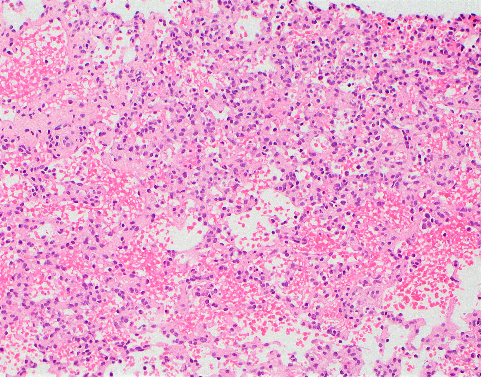
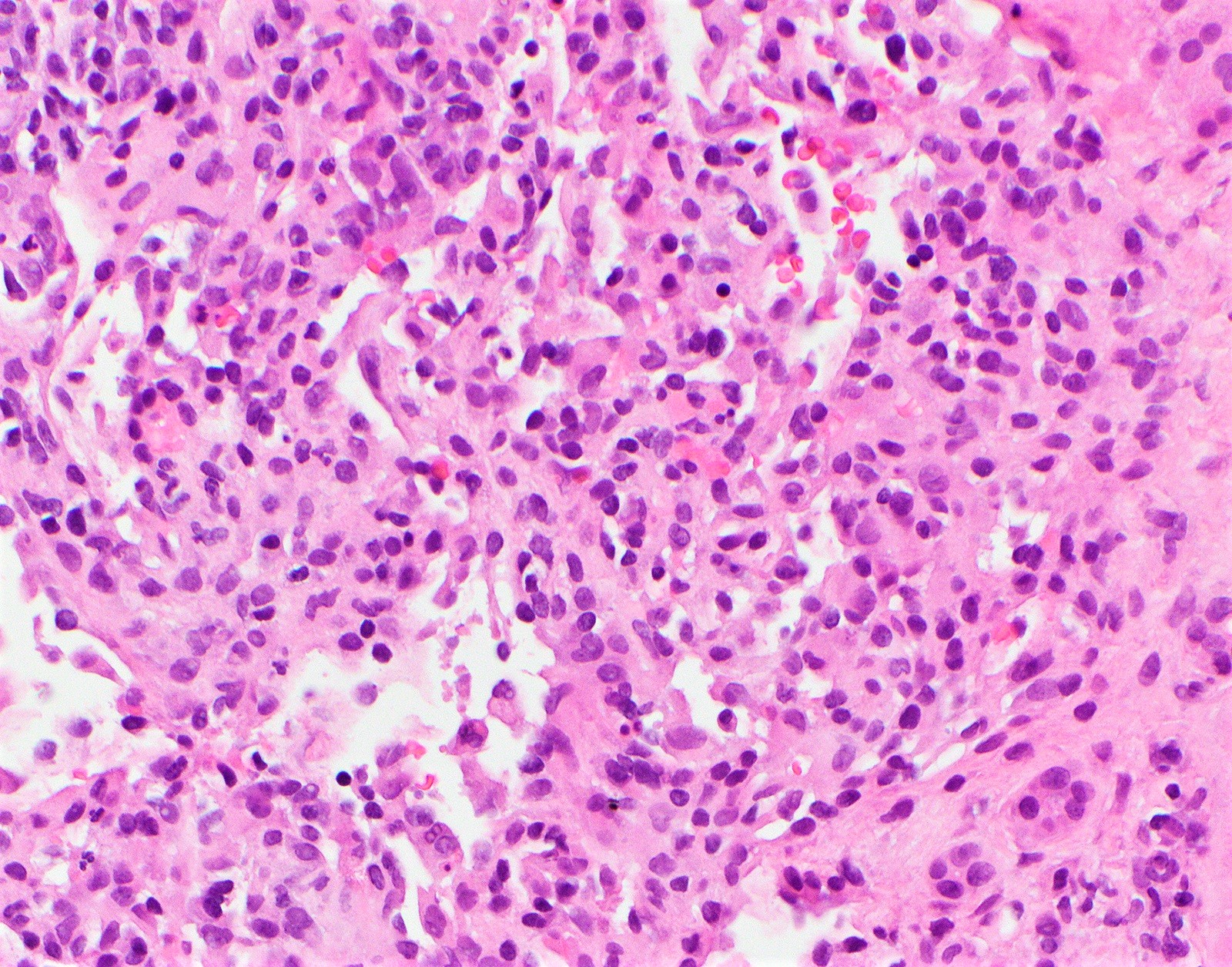
 Pyogenic Granuloma Lobular Capillary Hemangioma Histopathology Image 2 Path Quiz Case 70 Pathology Quiz Pathology Medical Science Anatomy And Physiology
Superficial on the surface of the skin deep under the skin and mixed.
Types of hemangioma pathology. Tumours of the lids. This type is much less common than IH. Some hemangiomas disappear over time and this is called regression.
But the main division into more and less dangerous conditions classification according to the degree of aggressiveness. A hemangioma is a common vascular birthmark made of extra blood vessels in the skin. This review highlights the key features of previously reported cases and discusses the differential diagnosis.
Hemangiomas with fluid-fluid levels. A congenital hemangioma can shrink on its own rapidly involuting or RICH or be nonshrinking noninvoluting or NICH. Congenital or infantile hemangiomas develop at or near birth.
A RICH will start to shrink right after the baby is born. Four histologic types of hemangioma have been identified. Several types are seen in soft tissue.
It is usually an incidental finding at autopsy but may present with abdominal pain. Doctors may diagnose a congenital hemangioma on a prenatal ultrasound. These features allow to separate vertebral hemangiomas from bone lymphangiomas from true neoplastic vascular tumours such as hemangioendotheliomas hemangiopericytomas angiosarcomas from hemangioblastomas and from arteriovenous malformations with shunt.
It is a benign non-cancerous growth. Often they disappear spontaneously but they. Numerous dilated thin walled vascular channels of variable size lined by flattened endothelial.
There are many types of hemangiomas and they can occur throughout the body including in skin muscle bone and internal organs. Results Of 472 hemangiomas 327 patients 339 72 were localized 84 18 were segmental 37 8 were indeterminate and 12 3 were multifocal 8 or more noncontiguous lesions. For cervical this pathology is even more dangerous.
1 2 According to the ISSVA classification hemangiomas are classified into infantile hemangiomas IH. Cavernous hemangioma is the most common primary hepatic tumor. A congenital hemangioma is present at birth and grows as a child grows.
This neurocutaneous syndrome is the most frequent one and it is associated with several types of vascular and non-vascular abnormalities which can involve any organ of the body. Other articles where Hemangioma is discussed. Neoplasm vascular anomaly is divided into several disorders which one of them is hemangioma.
Internal and external hemangiomas and hemangiomatous lesions progress and tend to regress concomitantly. Cystic or multilocular hemangiomas. A hemangioma is a benign noncancerous tumor made up of blood vessels.
Of the blood vessels called hemangiomas may occur in the lids and give rise to soft bluish swellings. A hemangioma is a common non-cancerous tumour made from blood vessels. Hemangiomas may occur anywhere on the body.
Cavernous Hemangioma of Liver. It is more common in females FM 51 in whom it may rapidly increase in size during pregnancy or with estrogen therapy. Subtypes were correlated with race and ethnicity the incidence of complications and overall outcome.
Most hemangiomas are infantile hemangiomas. Microscopic histologic description Two types. A congenital hemangioma hem-an-gee-o-ma is a vascular lesion that is present and fully grown at birth.
Rapidly involuting congenital hemangioma RICH and non-involuting congenital hemangioma NICH. A frequent type of atypical hepatic hemangioma is a lesion with an echoic border at ultrasonography. There are three main types of hemangioma.
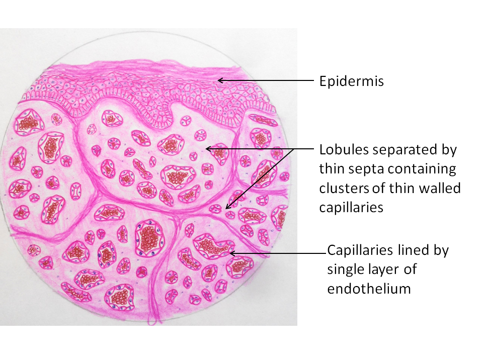
Less frequent types are large heterogeneous hemangiomas. Most hemangiomas occur on the surface of the skin or just beneath it. There are different kinds of hemangiomas and the most common types are called capillary cavernous and lobular capillary.
Before the year 2000 these lesions were grouped in with infantile hemangiomas. Congenital hemangiomas are usually divided into two groups. Cavernous and capillary Cavernous more common.
Capillarycavernous lobular capillary cellular and epithelioid. The most common hemangioma of the lumbar spine and a hemangioma of the thoracic.
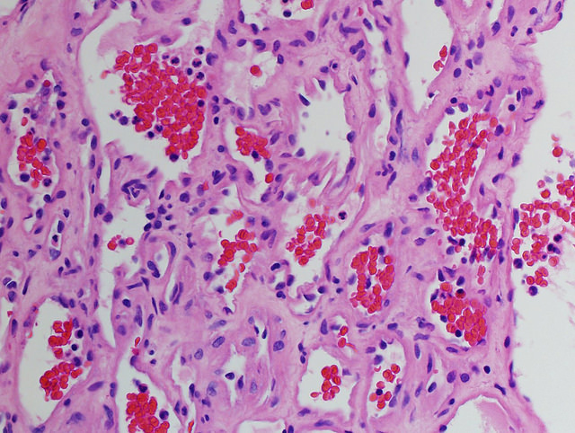
 Pathology Outlines Hemangioma
 Pathology Outlines Hemangioma
 Pathology Outlines Hemangioma
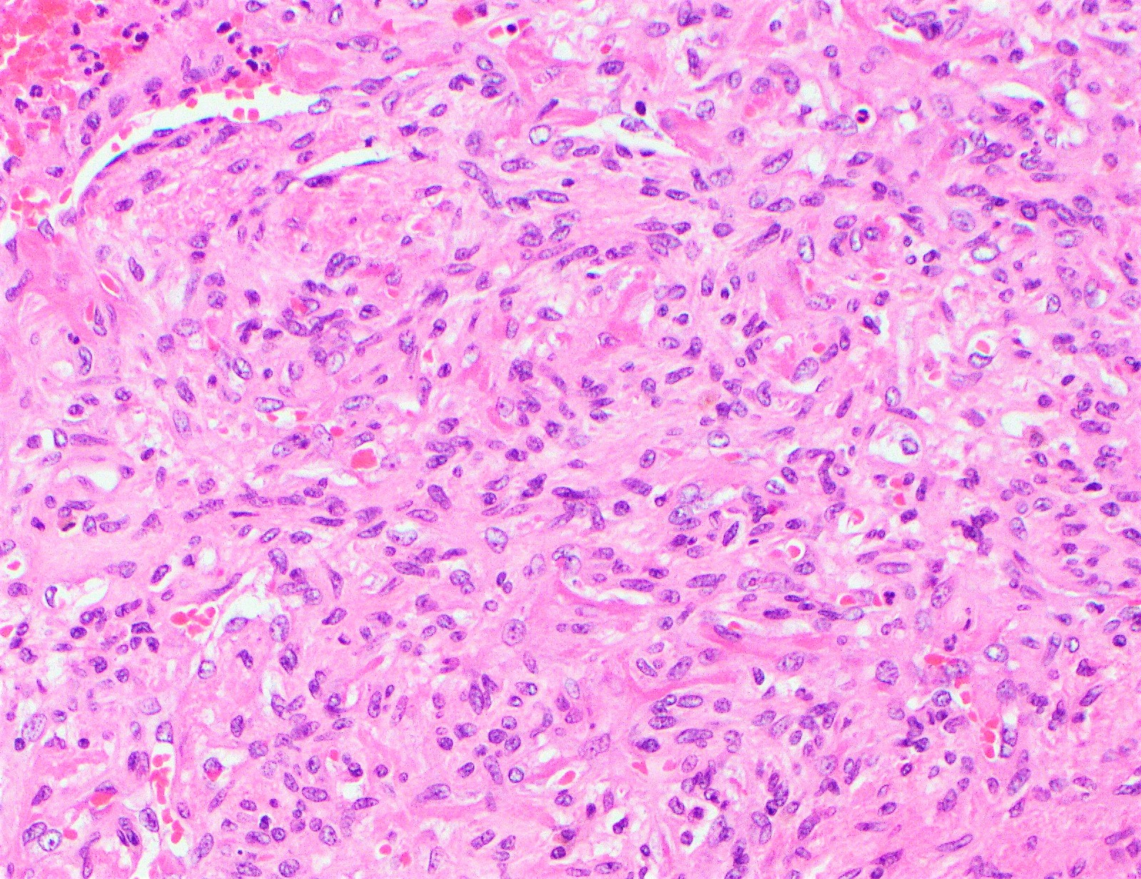
 Hemangioma Portnotes Orthopaedicsone
 Pathology Outlines Hemangioma
 Pathology Outlines Hemangioma
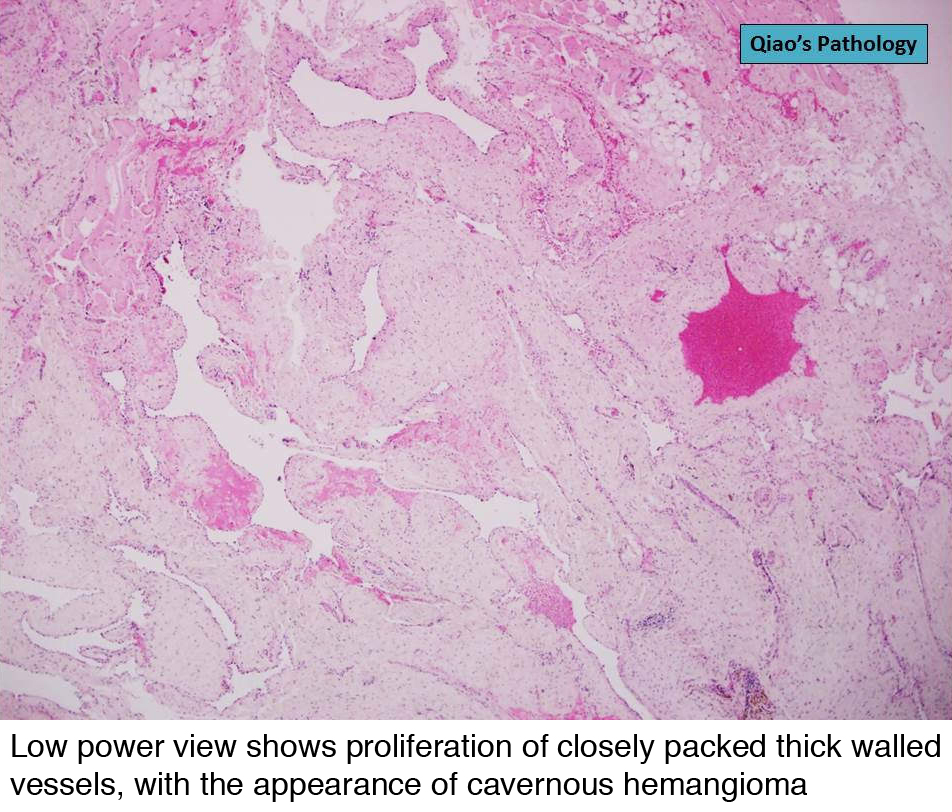
 Histology Of Mixed Cavernous And Capillary Angioma A Shows Cavernous Download Scientific Diagram
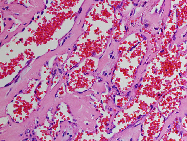
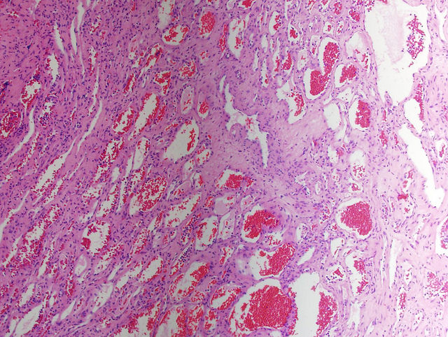
 Cavernous Hemangioma At 20x Magnification Nikon S Microscopyu
 Pathology Outlines Hemangioma
 Pathology Outlines Hemangioma

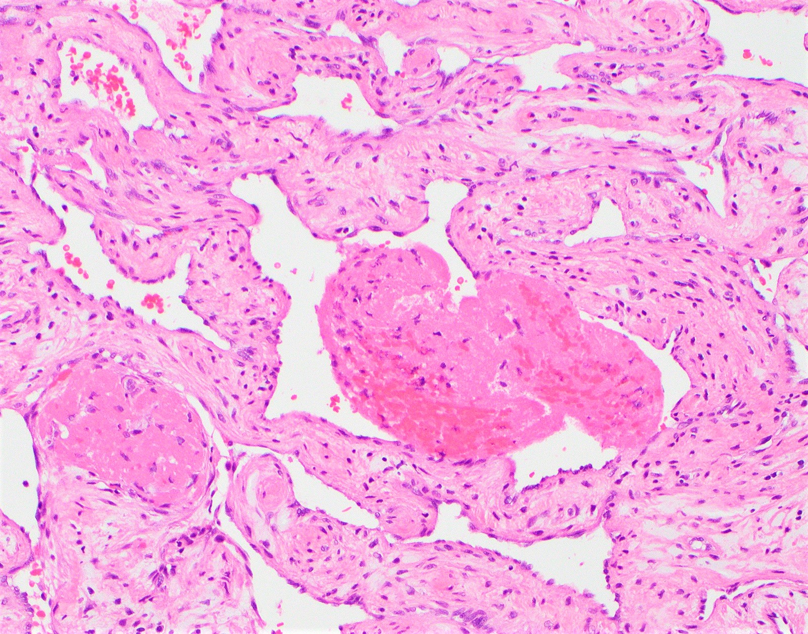
 Pathology Outlines Venous Hemangioma
 Lobular Capillary Hemangioma Mypathologyreport Ca

Pathology Outlines Hemangioma

 Histology A Cutaneous Hemangioma B Epithelioid Download Scientific Diagram